Okategoriserad

New Heartworks module with TAVR implant

Heartworks new module 1.4

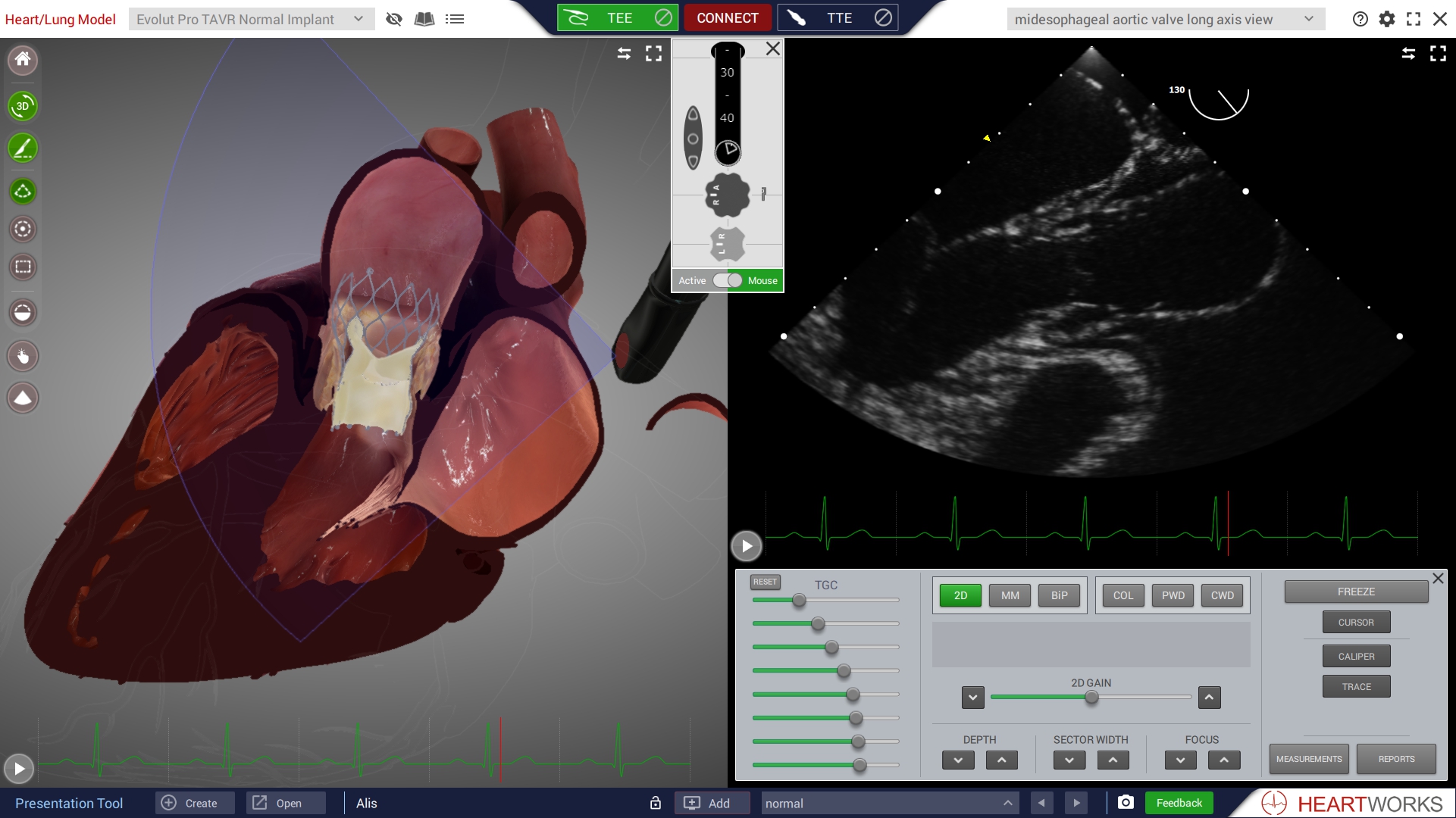
This release contains 5 examples of correct and incorrect TAVR placement, as well as touch screen capability for both screens, the functionality to turn off the rib shadows when scanning TTE, and important bug fixes.

TAVR devices are used in patients with severe aortic stenosis. The Medtronic Evolut PRO TAVR is a self-expanding transcatheter aortic valve deployed across the stenotic native aortic valve.

Evolut PRO TAVR Normal Implant

In this model the TAVR is correctly positioned. There is good flow through the valve and there are no paravalvular leaks, which can, and should, be confirmed using colour and spectral Doppler.

Evolut PRO TAVR Low Implant

n this model the TAVR is deployed in a ‘low’ position. The proximal end of the nitinol stent is positioned well below the native aortic valve annulus. This has resulted in a moderately severe paravalvular leak (PVL) in the right coronary sinus region of the native aortic root.

Evolut PRO TAVR High Implant

In this model the TAVR is deployed in a ‘high’ position. The proximal end of the nitinol stent is positioned above the native aortic valve annulus. This has resulted in a mild PVL near the commissure between the native left and non-coronary leaflets. This small jet runs immediately under the anterior leaflet of the mitral valve.

Evolut PRO TAVR Constrained Valve

In this model the TAVR is deployed in an appropriate position relative to the native aortic valve annulus but has failed to expand fully. A calcified native right coronary leaflet has prevented the valve reaching its fully expanded state. The cross-section of the valve stent is not circular and this has resulted in several areas of PVL. A large leak is present in the region close to the native non coronary sinus with another large leak near the commissure between the native left and right coronary leaflets.

Evolut PRO TAVR 80% Deployed

In this model the delivery system is seen passing through the native aortic valve with the TAVR stent approximately 80% deployed. The nose cone of the delivery system is seen in the LVOT and the spiral end of the Confida wire located in the left ventricle

Rib Shadows Filtering

Functionality to turn off the rib shadows when scanning TTE.